Our histological analysis provides a comprehensive assessment of biomaterial biocompatibility, tissue response, and bone regeneration following implantation. Using standardized staining techniques and immunohistology (IHC & IF), we characterize the host response and the integration of biomaterials in compliance with ISO 10993 guidelines.
Biocompatibility Assessment (ISO 10993)
We evaluate inflammatory response, fibrosis, and tissue integration through:
- Hematoxylin & Eosin (H&E): General tissue morphology and inflammatory cell infiltration.
- Masson’s Trichrome: Fibrosis and collagen deposition.
- Toluidine Blue/Safranin O: Cartilage integrity and glycosaminoglycan content.
- Immunohistology (IHC & IF): inflammation (CD68 for macrophage response).
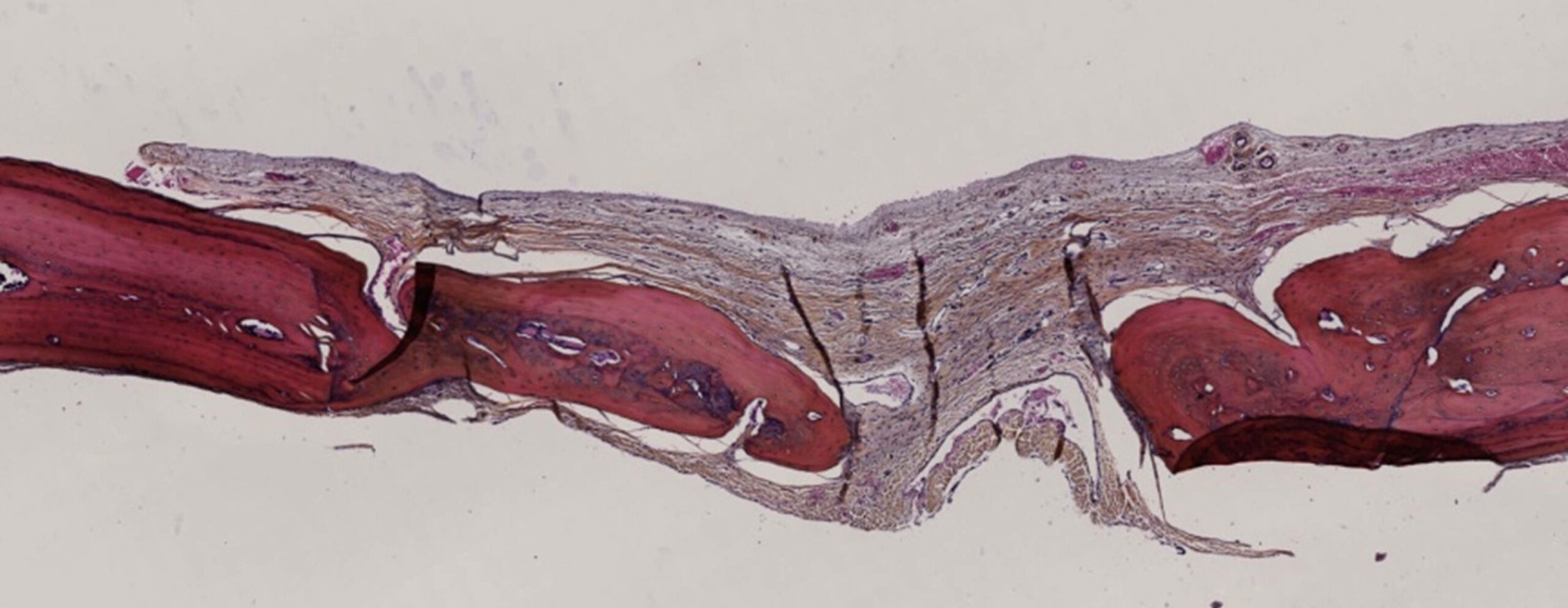
Bone Regeneration & Wound Healing Analysis
To assess bone healing and biomaterial osteoinduction, we use:
- Von Kossa: Mineralized bone matrix deposition.
- Goldner’s Trichrome: Differentiation between bone, osteoid, and connective tissue.
- Tartrate-Resistant Acid Phosphatase (TRAP): Osteoclast activity and bone remodeling.
- Immunohistology (IHC & IF): Osteogenic markers (e.g., osteopontin, osteocalcin), angiogenesis (CD31), and inflammation (CD68 for macrophage response).
Either resin embedding or paraffin embedding allows our team to work on markers adapted to the biomaterial being tested and/or the aim of the study.
Our high-resolution imaging and quantitative analysis enable precise evaluation of biomaterial performance, supporting implant development and tissue engineering research.