
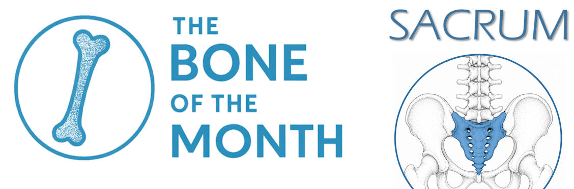
🦴 𝐁𝐨𝐧𝐞 𝐨𝐟 𝐭𝐡𝐞 𝐌𝐨𝐧𝐭𝐡 – 𝟕: 𝐓𝐡𝐞 𝐒𝐚𝐜𝐫𝐮𝐦 🦴
Exploring the fascinating world of bone biology…
Let’s continue our “𝐁𝐨𝐧𝐞 𝐨𝐟 𝐭𝐡𝐞 𝐌𝐨𝐧𝐭𝐡” series with a bone that literally sits at the center of our skeleton: the sacrum.
Located at the base of the spine, between the lumbar vertebrae and the pelvis, the sacrum is a triangular bone formed by the fusion of five vertebrae during adolescence and early adulthood.
Measuring approximately 10–12 cm in length, it acts as a crucial structural bridge, transferring the weight of the upper body to the pelvis and lower limbs while providing stability during standing, walking, and running.
𝐈𝐧 𝐩𝐫𝐞𝐜𝐥𝐢𝐧𝐢𝐜𝐚𝐥 𝐫𝐞𝐬𝐞𝐚𝐫𝐜𝐡, 𝐭𝐡𝐞 𝐬𝐚𝐜𝐫𝐮𝐦 𝐢𝐬 𝐫𝐞𝐥𝐞𝐯𝐚𝐧𝐭 𝐟𝐨𝐫:
✅ Understanding biomechanics and load transmission
✅ Studying bone remodeling under mechanical stress
✅ Investigating sacroiliac joint disorders and inflammation
✅ Evaluating regenerative therapies and biomaterials for bone repair
The sacrum is also closely linked to the sacroiliac joints, which connect the spine to the pelvis and play a fundamental role in posture, balance, and movement.
💡 Fun fact: Did you know women pelvis becomes slightly more flexible during pregnancy? Hormonal changes including the effects of relaxin, estrogen, and progesterone can increase the laxity of the ligaments around the pelvic joints. During childbirth, the sacrum and pelvic joints undergo subtle movements, allowing the pelvis to adapt slightly and helping facilitate the baby’s passage. It’s a remarkable example of the body’s natural adaptability.
At Atlantic Bone Screen, 𝐰𝐞 𝐞𝐱𝐩𝐥𝐨𝐫𝐞 𝐛𝐨𝐧𝐞 𝐛𝐢𝐨𝐥𝐨𝐠𝐲 𝐟𝐫𝐨𝐦 𝐦𝐢𝐜𝐫𝐨𝐬𝐭𝐫𝐮𝐜𝐭𝐮𝐫𝐞 𝐭𝐨 𝐟𝐮𝐧𝐜𝐭𝐢𝐨𝐧, supporting translational research and innovation in bone, dental, and osteoarticular diseases.
👀 Stay tuned for next month’s Bone of the Month and discover another fascinating structure from the human skeleton through the lens of science…