
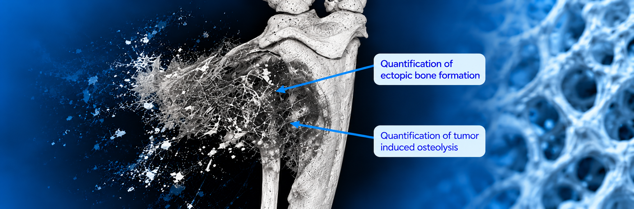
Bone Tumor Xenografts Profoundly Reshape Bone
Bone tumors do more than grow within or around bone, they can profoundly alter its architecture, density and remodeling dynamics.
How can we objectively characterize these structural changes?
Using high-resolution microCT, Atlantic Bone Screen provides a detailed 3D and quantitative assessment of tumor-bearing bones, enabling the evaluation of both bone destruction and abnormal bone formation.
Our analyses can include:
🔹 Tumor-induced osteolysis and bone destruction
🔹 Ectopic bone formation
🔹 Bone density: BV/TV, BMD, TMD
🔹 Trabecular architecture: Tb.N, Tb.Th, Tb.Sp
🔹 Cortical parameters: Ct.Th, Ct.Ar, porosity
🔹 3D reconstruction and segmentation to visualize and quantify structural changes
By combining relevant bone tumor xenograft models with advanced microCT analysis, we can turn complex tumor-induced bone remodeling into robust quantitative data.
And microCT is only part of the picture: these analyses can be complemented by histology, histomorphometry,IHC and IF to further characterize tumor–bone interactions at the tissue and cellular levels.
📩 Working on bone tumors or bone metastases? Let’s discuss how we can tailor the model and analysis strategy to your research objectives >
#BoneOncology #MicroCT #BoneMetastasis #Xenograft #BoneResearch #PreclinicalResearch #Histomorphometry #OncologyResearch