Radiography RX is used both during in vivo phases and for ex vivo analyses to visualize and analyse
Preclinical Imaging
Whether you require 3D analysis of bone architecture, high-resolution cellular imaging, or quantitative histological evaluations, our services are tailored to deliver reliable and actionable results.
To answer our Customers’ needs, Atlantic Bone Screen has developed a platform for image analyses with different techniques, microscopes and software. Our cutting-edge imaging capabilities provide detailed and quantitative insights into biological structures and processes. Combining state-of-the-art technologies with expert analysis, we support a wide range of preclinical studies with high-resolution data tailored to your research needs.
Most of the time the techniques give complementary information and the use of 2 or 3 methods in parallel consolidates the data and can help in the confirmation of the conclusion of a study.
Micro-computed tomography enables 3D reconstruction of bone samples for detailed architecture analyses, much more than BMD
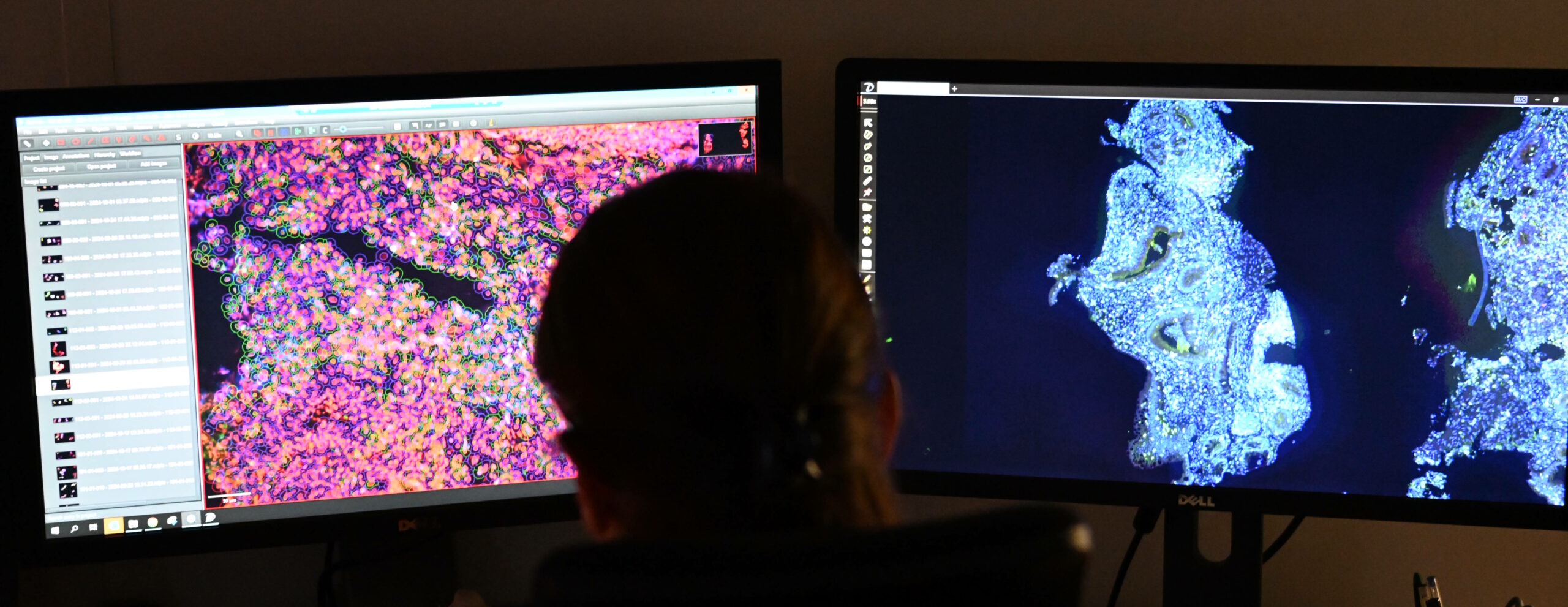
Both for cells and tissue visualization, we use our microscopes for precise analyses and descriptions
Comprehensive 2D & 3D Image Analysis for precise, data-rich results