Our bone and joint histology services provide a detailed analysis of bone remodeling, cartilage degradation, and inflammatory processes in preclinical models of osteoarthritis, osteoporosis, and other musculoskeletal diseases. We combine classical histology and immunohistochemistry (IHC), and also 3D microCT analyses to deliver a comprehensive evaluation of bone and joint pathology.
Bone Histology & Remodeling Analysis
To assess bone formation, resorption, and remodeling, we use:
- Goldner’s Trichrome: Differentiation between mineralized bone, osteoid, and connective tissue.
- Von Kossa/Alizarin Red: Bone mineralization and calcium deposition.
- Tartrate-Resistant Acid Phosphatase (TRAP): Osteoclast activity and bone resorption.
- Immunohistochemistry (IHC): Osteoblast (osteopontin, osteocalcin) and osteoclast (cathepsin K) markers.
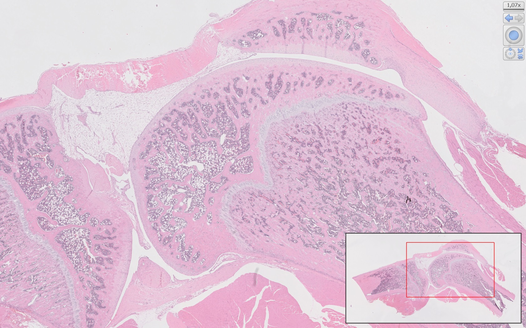
Cartilage & Joint Tissue Evaluation
For osteoarthritis and joint degeneration studies, we provide:
- Hematoxylin & Eosin (H&E): General cartilage and synovial tissue morphology.
- Safranin O/Fast Green: Glycosaminoglycan content and cartilage matrix integrity.
- Toluidine Blue: Proteoglycan distribution in articular cartilage.
- Immunohistochemistry (IHC): Cartilage turnover (collagen type II, aggrecan), inflammation (TNF-α, IL-1β), and chondrocyte activity (MMP-13).
MicroCT used as a Complementary Tool for Bone & Joint Analysis
To complement histological evaluation, our microCT imaging provides high-resolution insights into bone volume, microarchitecture, and trabecular structure / subchondral bone changes in osteoarthritis models / Osteophyte formation and joint space narrowing.
By combining quantitative microCT and histological analysis, we ensure a comprehensive evaluation of bone and joint health, supporting the development of therapeutic strategies for musculoskeletal disorders.