Our histology services provide in-depth analysis of tumor architecture, cell proliferation, and angiogenesis using gold-standard staining techniques and advanced immunostainings (IHC/IF).
Sample Preparation
Tumor biopsies are processed through resin or paraffin embedding, or frozen sections. Any kind of biopsies can be processed and received either in formol or already embedded.
Staining Techniques & Tumor Microenvironment Analysis
We perform classical histological stains, including:
- Hematoxylin & Eosin (H&E): general tissue morphology evaluation and tumor architecture
- Masson’s Trichrome (MT): for collagen fibers highlighting fibrosis and extracellular matrix characterization.
- Periodic Acid-Schiff (PAS): carbohydrates, glycoproteins and basement membrane integrity.
Other specific stain can be applied to highlight specific tissue components under request, depending on tissue and studied model and cancer.
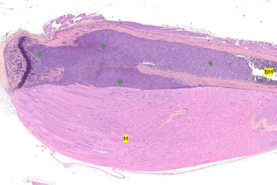
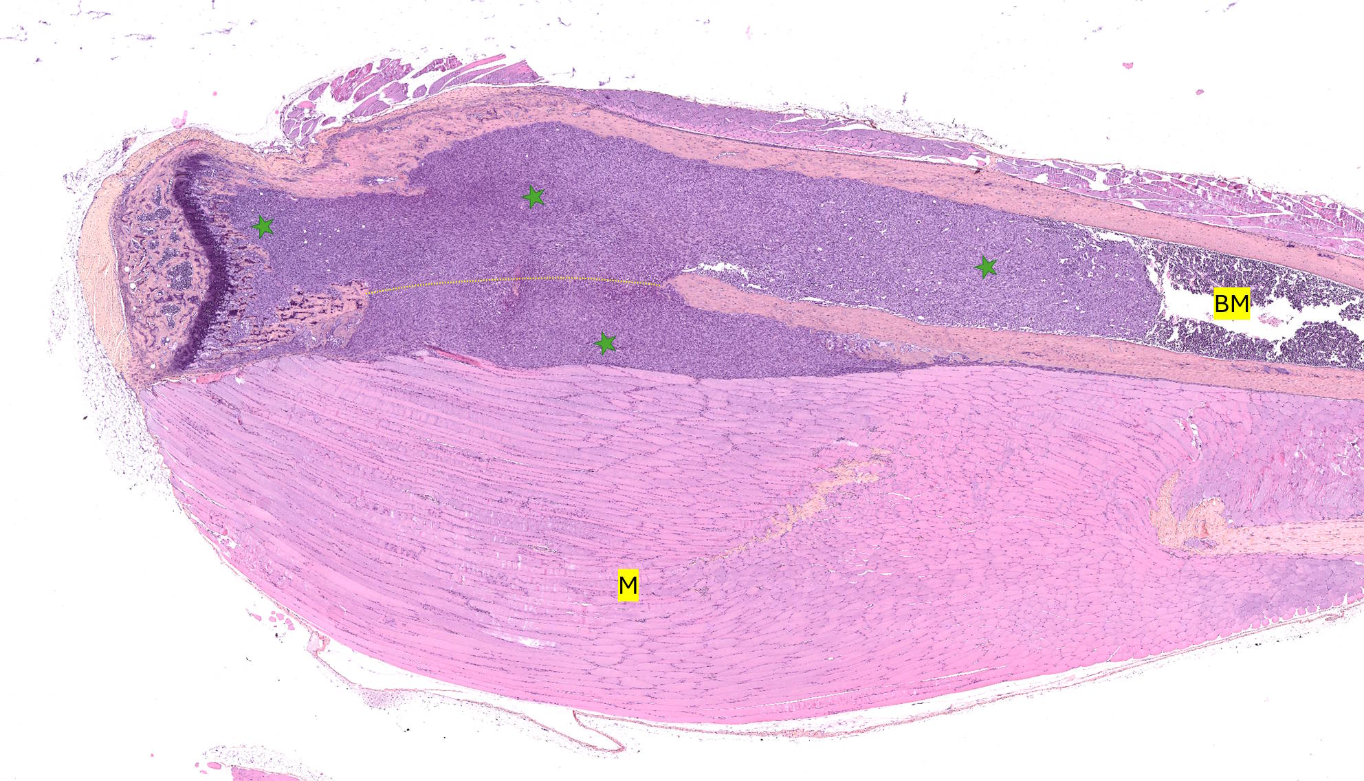
Histological image (HES stained) of murine tibias 28 days after intratibial injection of breast cancer cells. Tumoral development (green star) and invasion is visualised throughout the entire bone cavity. Yellow dotted line represents the degraded cortical bone due to tumoral development. M: muscle, BM: bone marrow
Immunohistochemistry (IHC) and Immunofluorescence (IF) for Tumor Characterization
Used to detect, identify and quantify specific tumor-associated proteins and other biomarkers. Both IHC and IF protocols can be customized for specific research studies, including investigations into new anti-cancer treatments.
As examples to assess key oncological markers, we can especially work on:
- Cell proliferation: Ki-67
- Angiogenesis: CD31
- Apoptosis & necrosis: TUNEL assay
- Tumor microenvironment: Immune cell infiltration (CD3, CD8, F4/80).
Histomorphometric analyses
Quantitative evaluation of tumor architecture, angiogenesis (new blood vessel formation), and cell proliferation.
Histopathological evaluation
Detailed histopathological assessments provide insights into tumor behavior, aiding in mechanism-of-action studies and therapeutic development.
Our preclinical imaging and quantitative analysis thus support accurate assessment of tumor progression and therapeutic efficacy.