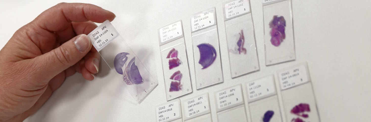
Histology for Toxicology (Non-GLP)
Our toxicological histology services provide a comprehensive evaluation of tissue integrity, cellular damage, and inflammatory responses across various organs and species. Conducted in a non-GLP (Good Laboratory Practice) environment, our analyses support early-stage preclinical research, safety assessments, and proof-of-concept studies. Both samples generated on our platform and samples we receive in formol or embedded in paraffin can be processed.
Comprehensive Organ Histology
We analyze a wide range of organs to detect histopathological changes, including:
- Liver: Hepatocellular degeneration, steatosis, fibrosis
- Kidney: Tubular damage, glomerular alterations, fibrosis
- Heart: Myocardial necrosis, fibrosis, vascular integrity
- Lungs: Inflammation, fibrosis, alveolar damage
- Spleen & Lymph Nodes: Immune response, hyperplasia, atrophy
Standard stainings and immunostaining (IHC & IF) are available as routine protocols: H&E / HES, Masson’s Trichrome, PAS, Siruis Red… And we can also develop new protocols under request.
Toxicological Biomarkers & Immunohistology
We perform immunohistological staining (IHC-IF) to detect key toxicological biomarkers, including:
- Apoptosis & Cell Death: Caspase-3, TUNEL assay.
- Oxidative Stress & Inflammation: COX-2, TNF-α, IL-6.
- Fibrosis & Tissue Remodeling: α-SMA, Collagen I/III.
- Vascular Integrity & Angiogenesis: CD31.
Species & Study Adaptability
Our expertise spans rodents (mice, rats), rabbits, and large animal models, allowing flexible adaptation to diverse preclinical toxicology studies.
Flexibility & Customization
As a non-GLP service, we offer tailored protocols, rapid study execution, and exploratory analyses, ensuring valuable insights for early-stage research and decision-making in drug development.