
3D MicroCT
Micro-CT Imaging for Bone Analysis
Our micro-computed tomography (microCT) service provides high-resolution 3D imaging for precise, non-destructive analysis of bone structure. This advanced technique is ideal for:
- Quantitative evaluation of trabecular and cortical bone parameters
- High-resolution imaging to study bone remodeling, biomaterial integration, and fracture healing
- Longitudinal studies, enabling repeated scans over time and preserving samples for complementary analyses (e.g., histology, biomechanics)
MicroCT Workflow
- Acquisition: High-resolution imaging using the SkyScan system
- Reconstruction: Generation of 2D binarized cross-sections from raw data
- Analysis: Qualitative and quantitative assessment on virtual slices in all spatial planes
MicroCT is particularly suited for evaluating bone loss (e.g., osteoporosis models) and comparing bone parameters such as Bone Volume Fraction (BVF), Bone Mineral Density (BMD), Trabecular Thickness (Tb.Th), Number (Tb.N), and Separation (Tb.Sp), across experimental groups. It’s a powerful tool to visualize and quantify the impact of compounds, biomaterials, or nutraceuticals on bone architecture.
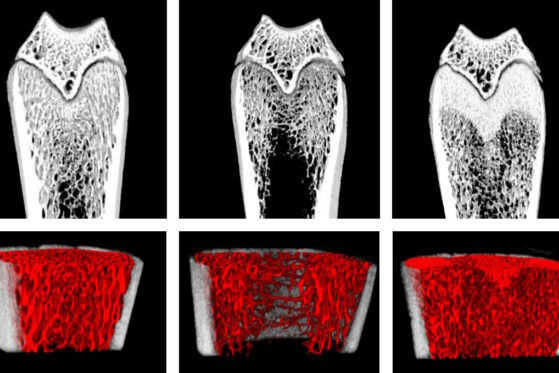 Example: Osteoporosis model induced by ovariectomy