Microscopy preclinical imaging
Microscopy for cells visualisation and for histology
We offer a wide range of microscopy modalities to support in vitro and histological studies:
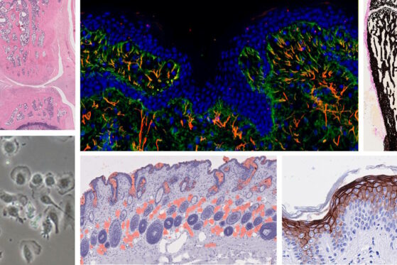
- Conventional microscopy (brightfield and fluorescence) for routine histology and structural analyses, and for cells visualization during in vitro cultures to evaluate cell differentiation.
- Slide scanner for high-throughput, whole-slide imaging system (brightfield and fluorescence) and high-quality digitalization of stained or immulolabelled (IF, IHC) tissue.
- Confocal microscopy for high-resolution 3D imaging of cellular structures and histological sections.
These techniques allow us to visualize cellular morphology, protein localization, and tissue architecture.
High-quality generated images are then analysed by automatic and IA assisted solutions.