Atlantic Bone Screen can evaluate the efficacy and cytotoxicity of compounds on tumoral cell lines from human or animal sources
Oncological models
Assess the effect of your compounds on bone cancer and metastases
Bone cancer can be classified into two main types: primary and secondary cancer. While both can affect bones, they differ in origin, progression and treatment.
Primary bone cancer begins in the bone itself and is relatively rare. The most common types include osteosarcoma, chondrosarcoma, and Ewing sarcoma. Secondary bone cancer, also known as bone metastases, occurs when cancer from another part of the body – such as the breast, lungs, or prostate – spreads to the bones.
To mimic tumoral development and evaluate compounds, tumoral cells can be implanted by subcutaneous injections in immunocompromised animals (xenograft or PDX), or directly nearby bone with paratibial or intratibial implantation. Different cell types have been used by Atlantic Bone Screen, such as MDAMB 231 (human breast cancer cell line) or OSRGa (rat osteosarcoma cell line).
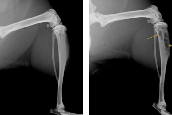
Xray images of murine tibias. Left: healthy tibia – right: after intra tibial injection of breast cancer cells. Yellow arrows represent areas of bone resorption, linked to tumor development
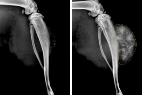
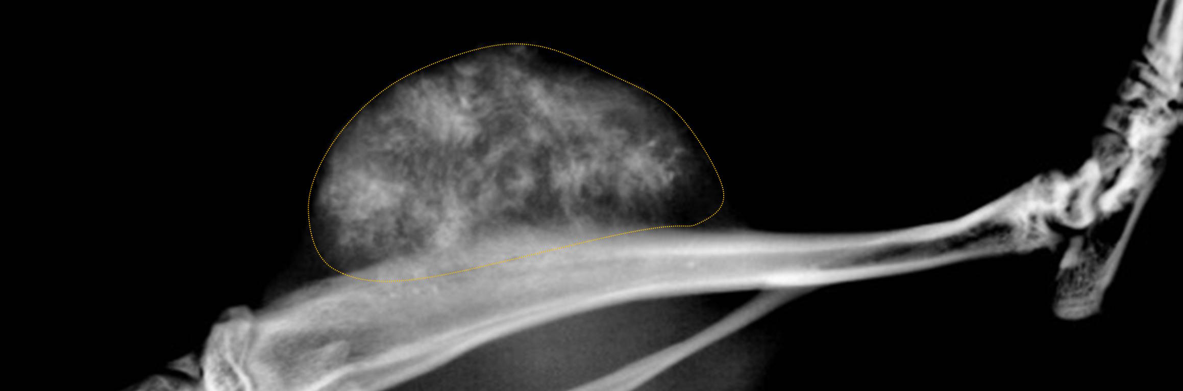
Xray images of rat tibias. Left: healthy tibia – right: after paratibial implantation of osteosarcoma tumoral cells. Yellow dotted lines represent ectopic bone formation, linked to tumor development
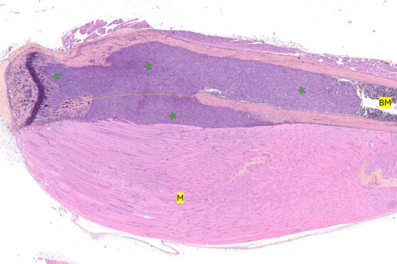
Histological image (HES stained) of murine tibias 28 days after intratibial injection of breast cancer cells. Tumoral development (green star) and invasion is visualised throughout the entire bone cavity. Yellow dotted line represents the degraded cortical bone due to tumoral development. M: muscle, BM: bone marrow
Expert services for histology in oncology involve advanced techniques used to analyze tumor tissues, their microenvironment, metastasis presence...
Thanks to current capabilities and expertise, our Team can develop animal models for the assessment of your compounds and biomaterials