Assess the effect of your compounds on osteoporotic animals model
Thanks to its expertise in bone pathologies, Atlantic Bone Screen provides preclinical animal models miming human bone pathologies to test drug candidates and nutraceuticals.
Among these bone pathologies, post-menopausal osteoporosis is a pathology resulting from an imbalance between bone formation and bone resorption, favoring bone resorption and resulting in a loss of bone mass and structural disorders.
After ovariectomy of mice or rats, bone resorption rate will exceed bone formation rate and resulting in a bone loss and an osteoporotic phenotype. Post-ovariectomy lesional modifications are detected (on the rats) on proximal metaphysis of long bones. Bisphosphonates, generally indicated for osteoporosis treatment, does not give full satisfaction as far as they have disabling secondary effects; alternatives to this treatment are thus necessary and a lot of Companies are working on new molecules for new optimized treatments.
Reference compound used is for example Alendronate, an antiresorptive agent belonging to bisphosphonate familly widely used to treat osteoporosis since it increases bone mineral density, decreases bone turnover, and reduces the risk of vertebral fracture among women with osteoporosis.
MicroCT analyses, bone biomarkers (such as PiNP, CTX1) and histology (HE, Von Kossa, TRAP staining …) followed by static or dynamic histomorphometry and histopathological analyses are the main read-outs to evaluate the efficacy of the tested drug candidates and compare data from a group to another one.
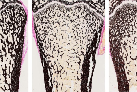
Representative images of Von Kossa staining on slides from resin embedded rat femurs. Left: femur of healthy rat, middle: femur of ovariectomised rat (with osteoporosis) and right: ovariectomised rat treated with alendronate, a bisphosphonate preventing bone resorption. Mineralised bone appears in black, collagen and connective tissue appear in red.
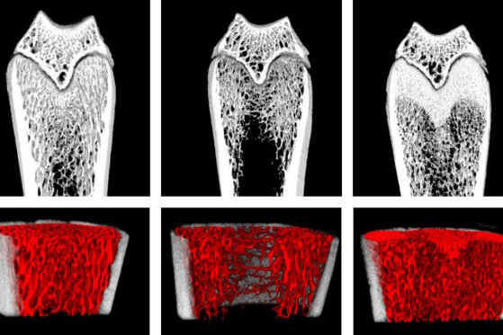
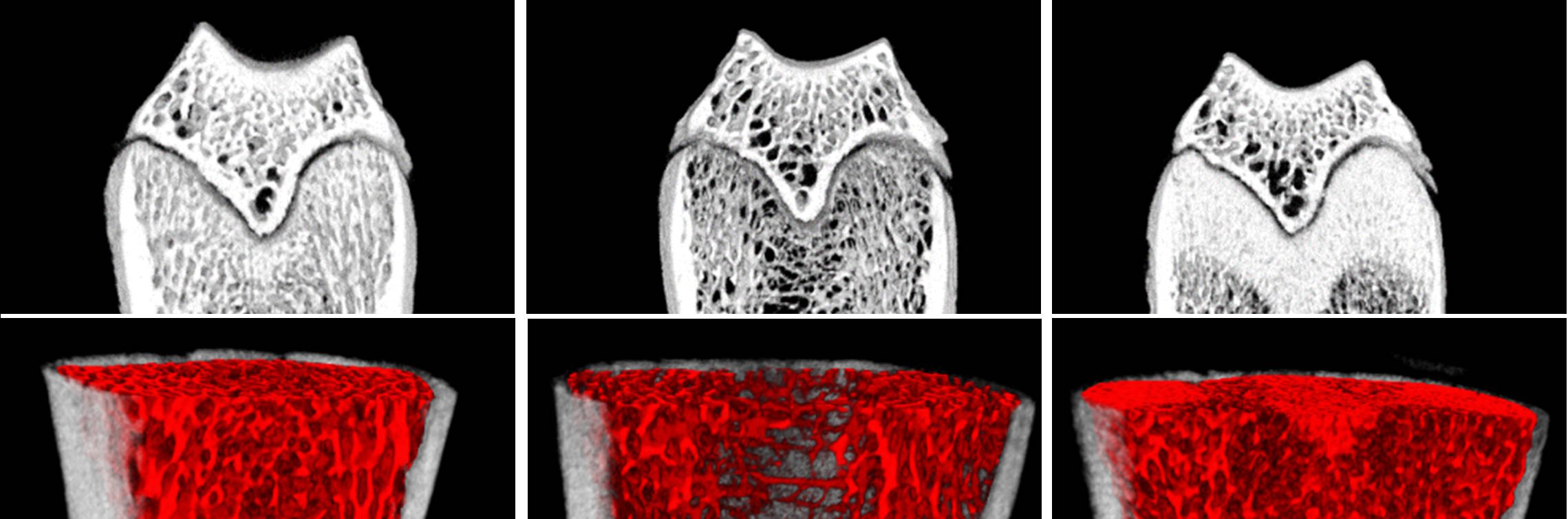
Representative images of micro-CT acquisition of rat femur, 8 weeks after surgery. Images of healthy rat, ovariectomised rat (with osteoporosis) and ovariectomised rat treated with alendronate, a bisphosphonate preventing bone resorption. Upper panel: 3D reconstruction of femur, coronal view; Lower panel: 3D reconstruction of trabecular bone, highlighted in red. Cortical bone appears in white.