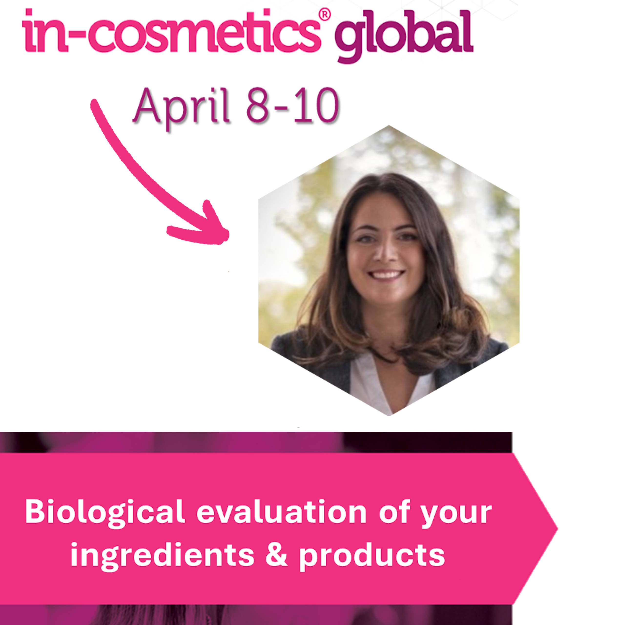
Meet us during InCosmetics in Amsterdam 04.2025 Meet us during In-Cosmetics 2025 🫧 𝐋𝐞𝐭’𝐬 𝐜𝐨𝐧𝐧𝐞𝐜𝐭 𝐝𝐮𝐫𝐢𝐧𝐠 𝐢𝐧-𝐜𝐨𝐬𝐦𝐞𝐭𝐢𝐜𝐬 𝐧𝐞𝐱𝐭 𝐰𝐞𝐞𝐤 𝐢𝐧 𝐀𝐦𝐬𝐭𝐞𝐫𝐝𝐚𝐦! 🫧 We’re excited to attend this
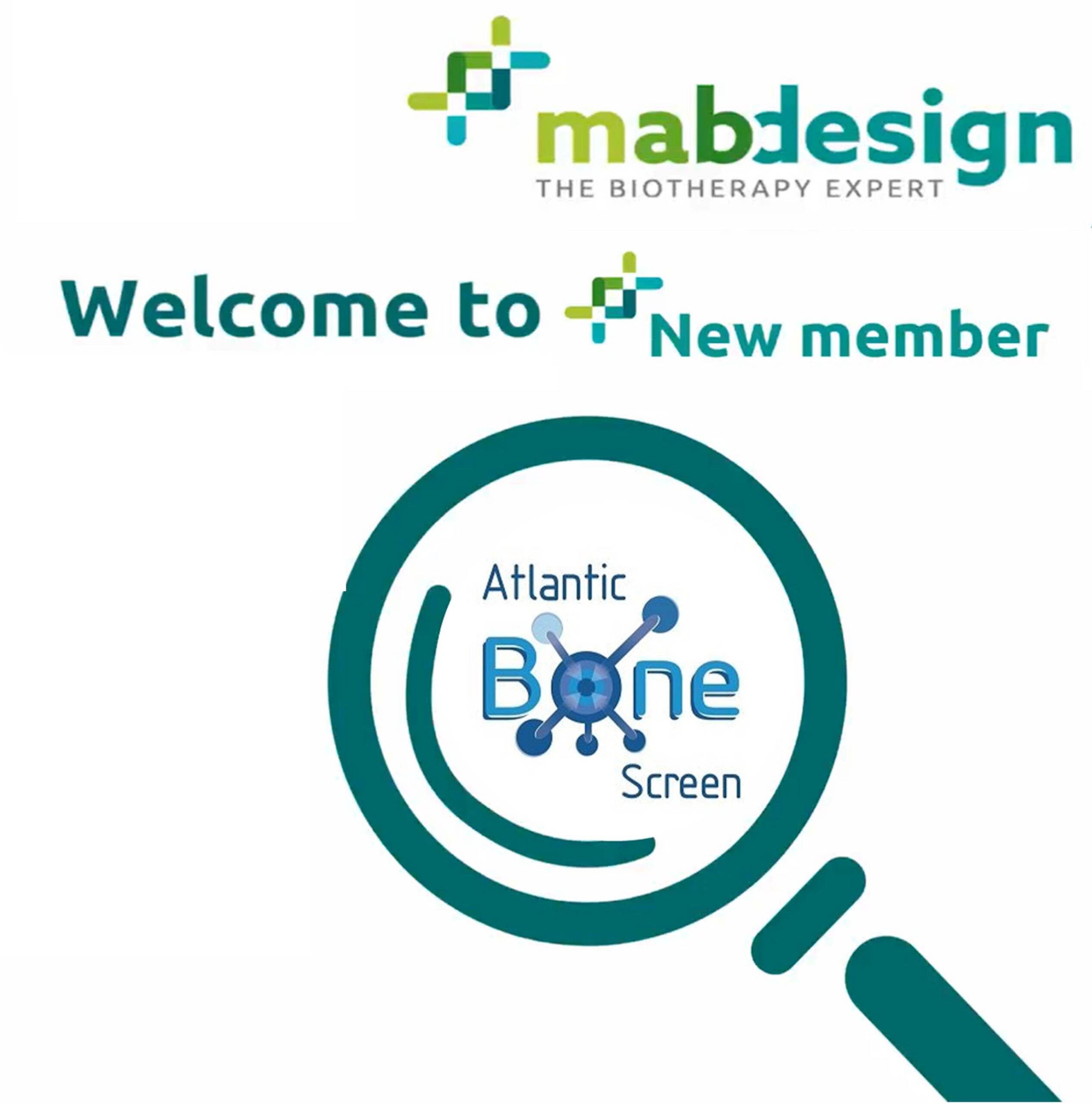
Member of Mabdesign for 2025 03.2025 Proud New member of Mabdesign Association 🎉 We are excited to announce that we are now a proud
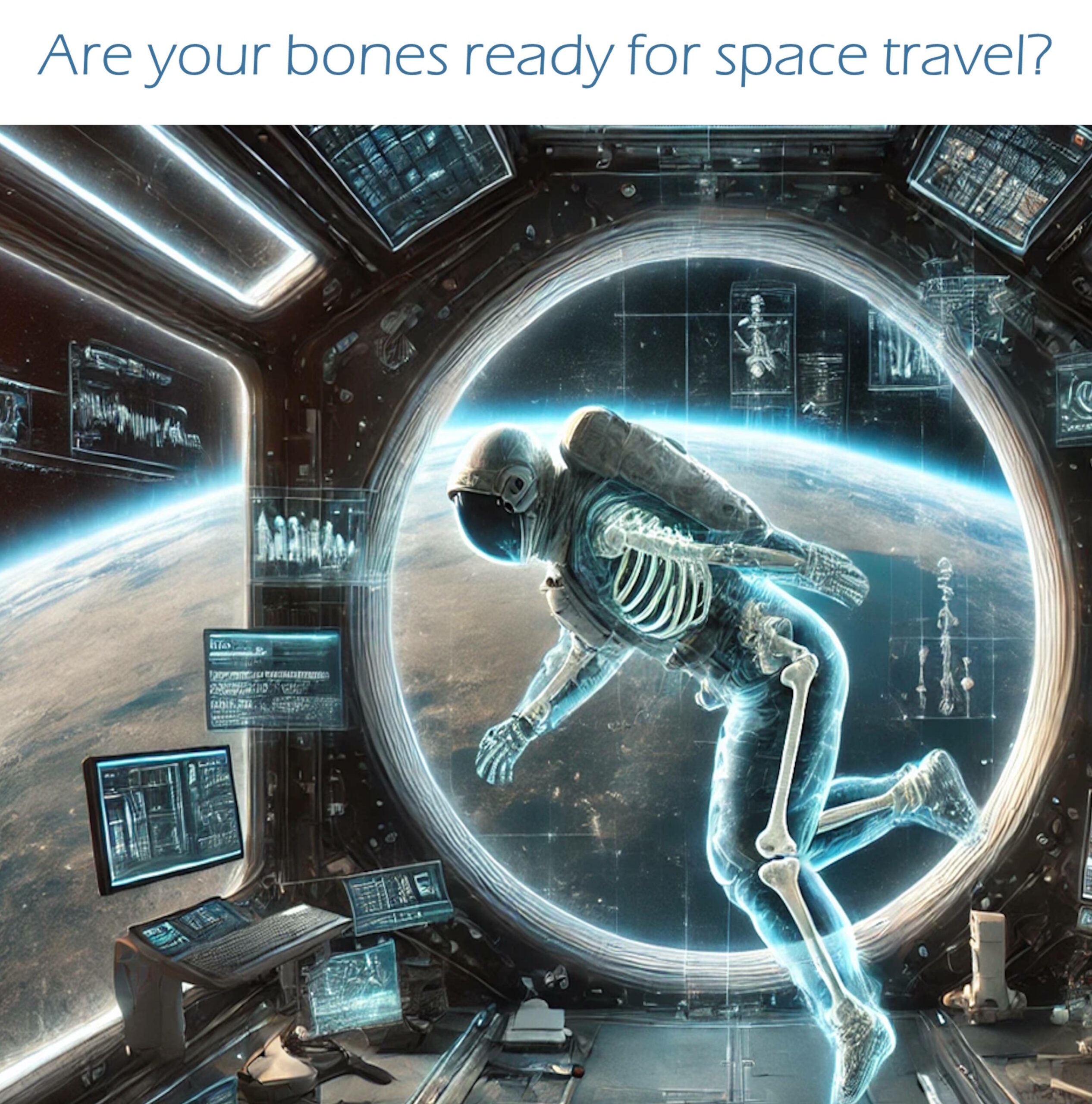
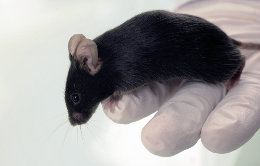
Are your bones ready for space travel? 03.2025 Are your bones ready for space travel? Bone Health & Space travel: What happens after months in microgravity?
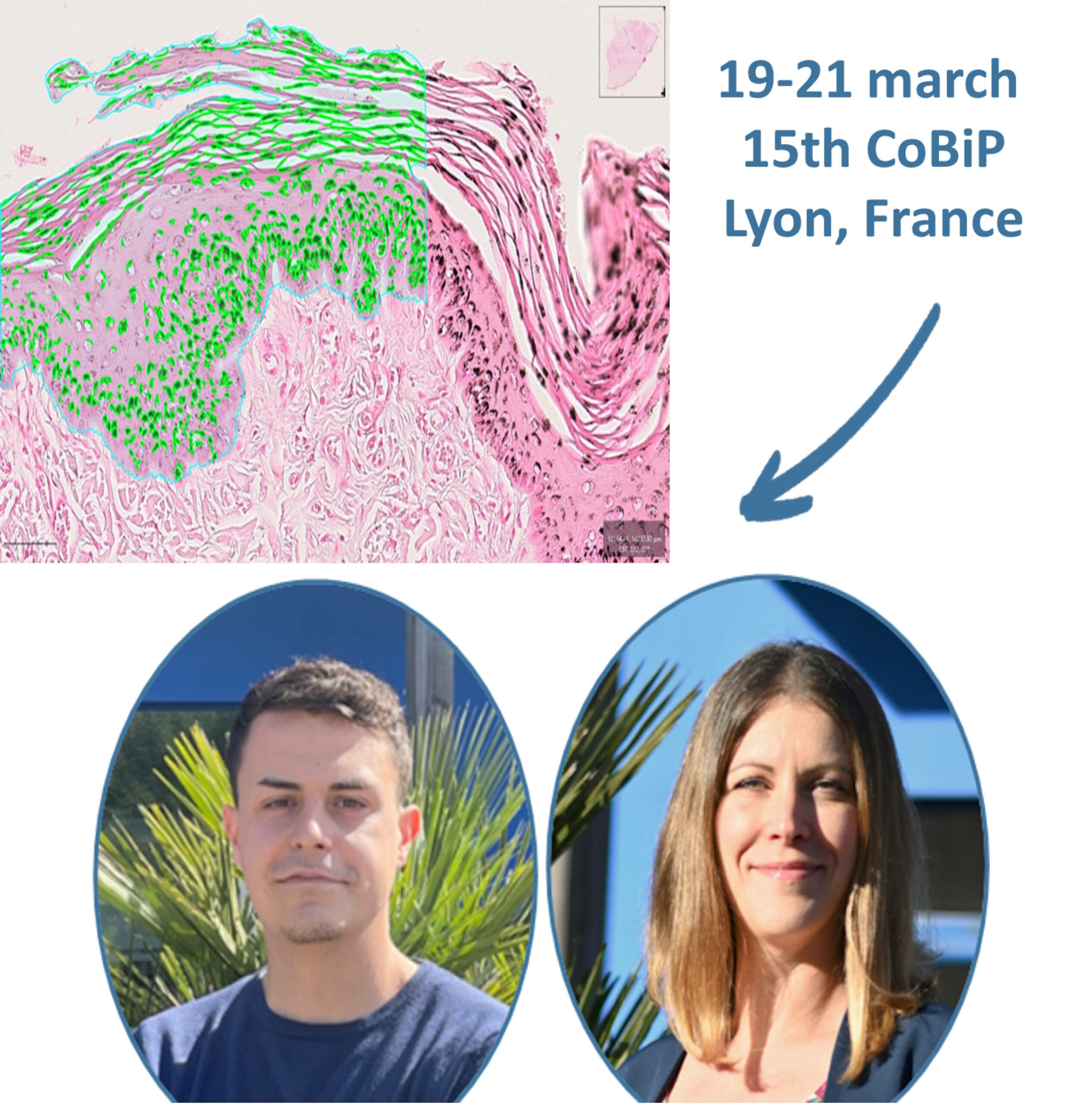
Meet us in CoBiP 2025 in Lyon 03.2025 Meet us in CoBiP in Lyon 📢On March 19th, 20th and 21st, our Team will be participating 15th
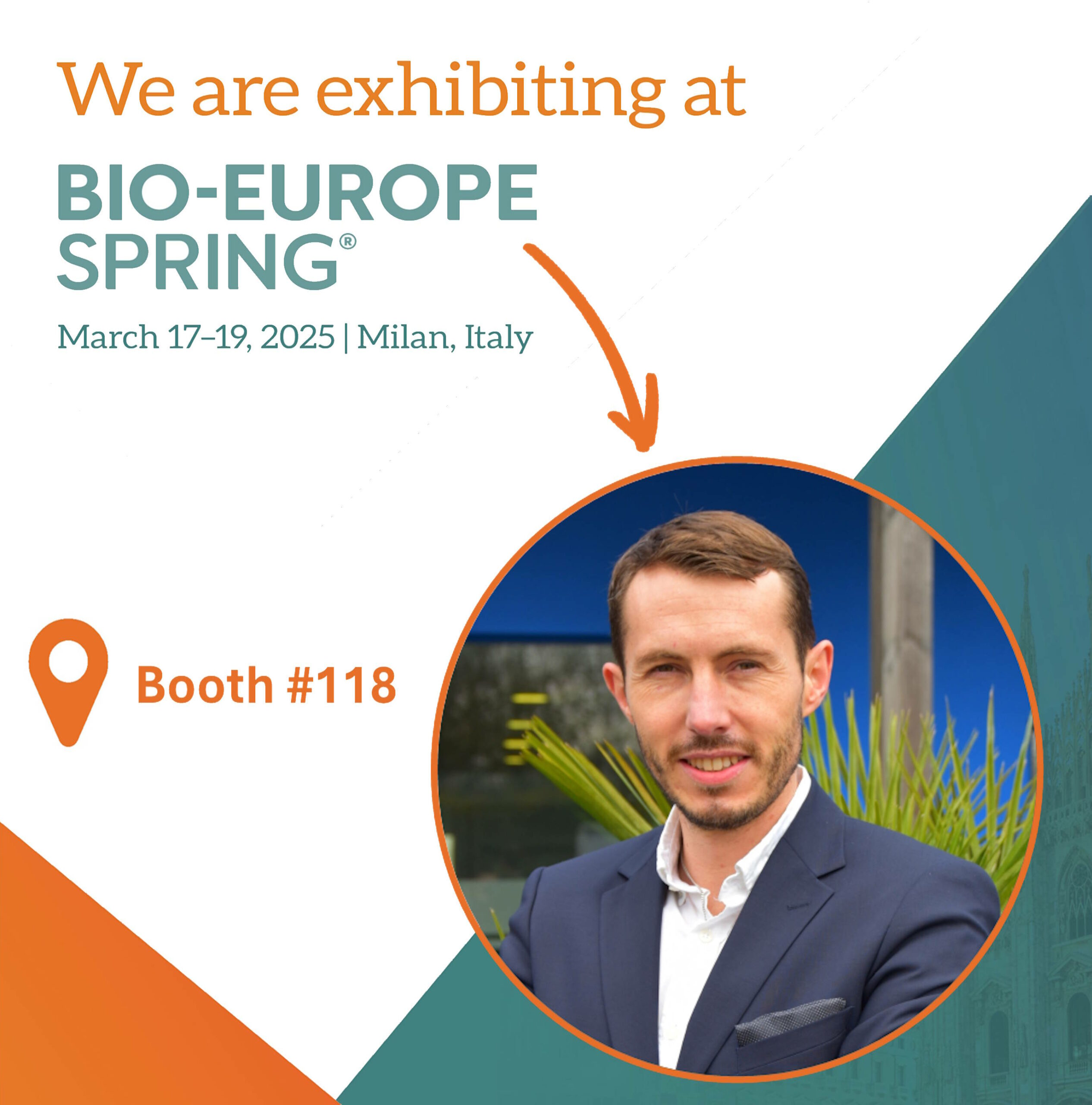
Meet us in Bioeurope in Milan 02.2025 Meet us in BioEurope in Milan We are heading to BioEurope Spring 2025 in Milan! We are excited
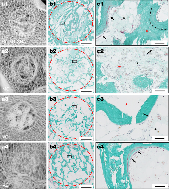
Mineralized collagen plywood contributes to bone autograft performance 12.2024 Autologous bone (AB) is the gold standard for bone-replacement surgeries1, despite its limited availability and the need for
Atlantic Bone Screen 12.2024 Optimize and accelerate the development of your compounds. High-Quality expertise in the field of bone and joint diseases,
Meet us in Biofit in Lille 11.2024 Meet us in Biofit in Lille We are thrilled to announce our participation next week in the 2024
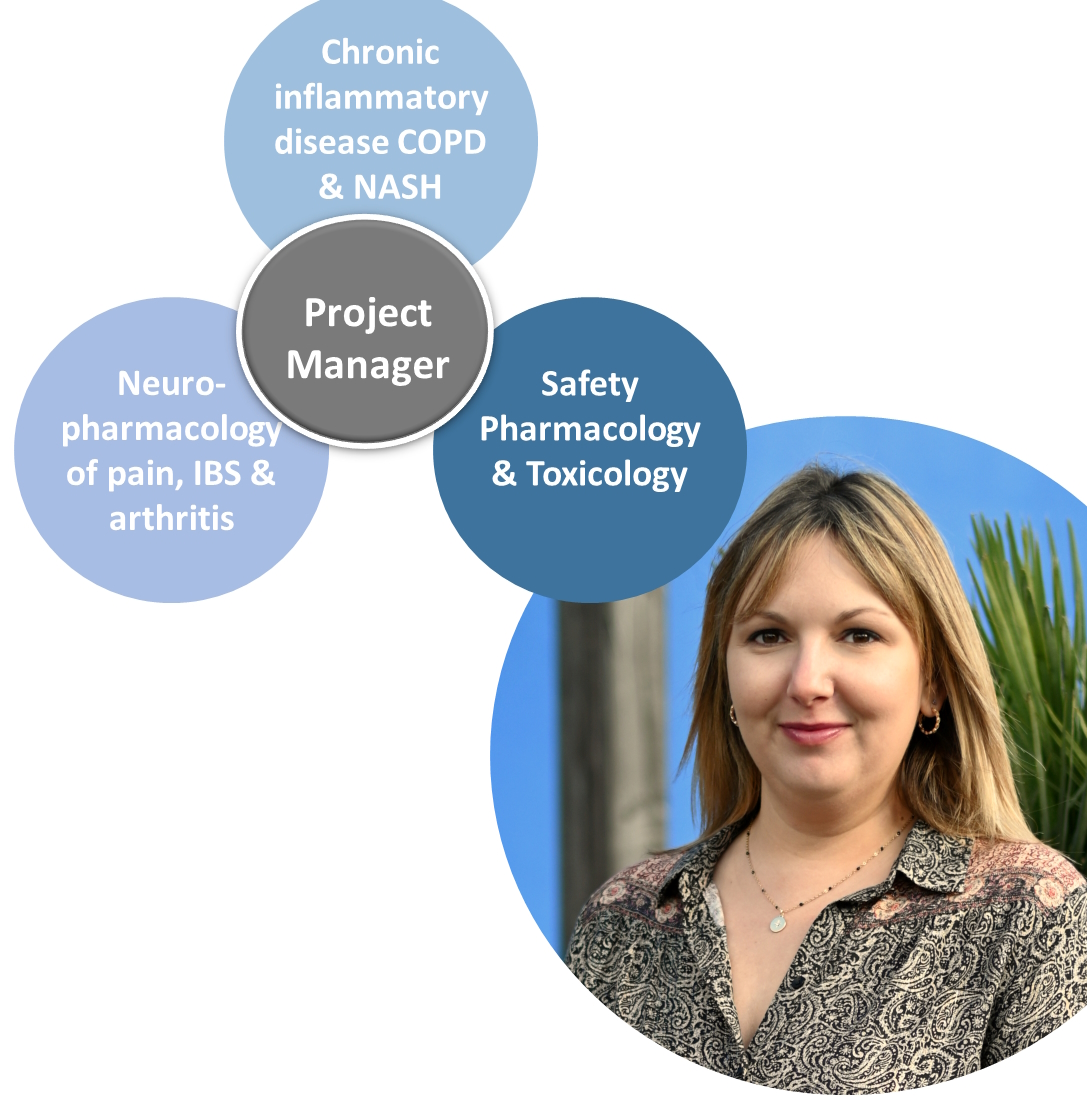
Lights on… Elodie Picard, PhD 10.2024 Lights on … Elodie Picard, PhD, Project Manager ⭐⭐Lumière sur…. Elodie Picard, PhD, Project Manager at ATLANTIC BONE
Tailor-made histology and imaging analysis 09.2024 Histological and imagery services as an endpoint for in vivo analysis Ex-vivo analysis of your own samples Download
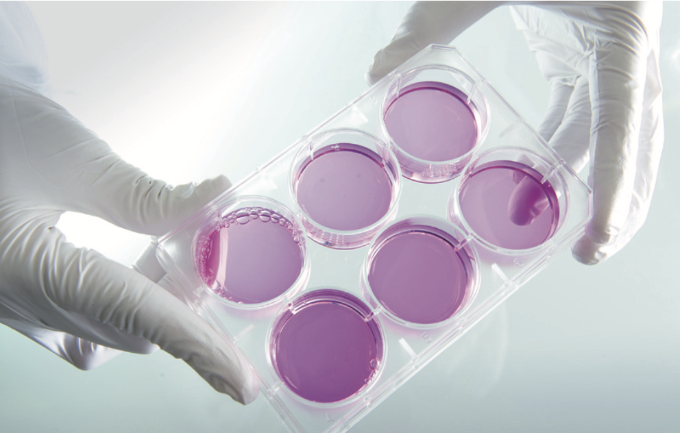
High-Quality in vitro assays for the screening and evaluation of your compounds 09.2024 Applications for Health Care, Food and Cosmetic Industries : Drug candidates, Nutraceutials, Biomaterials, Formulations and more. Primary cells
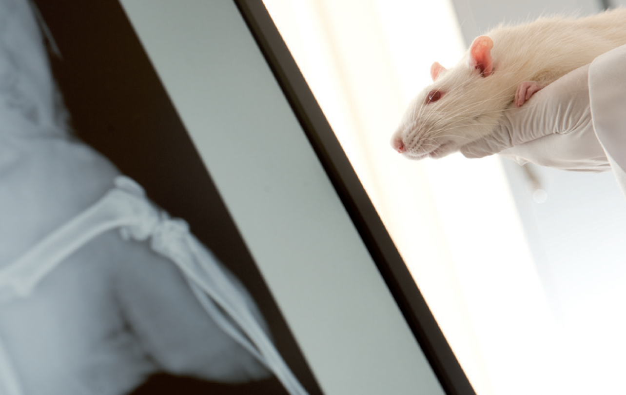
Reliable animal models reproducing human bone pathologies 09.2024 Identification and characterization of the therapeutic effect of your compounds. Applications for Health Care, Food and Cosmetic Industries